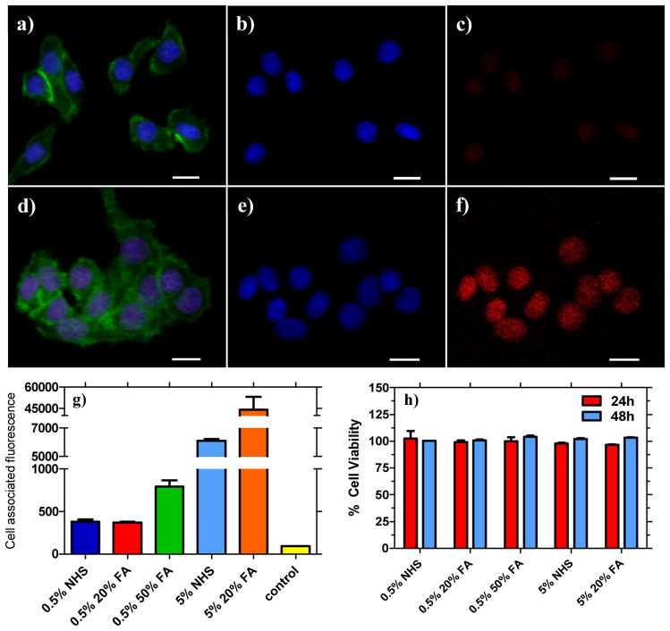
Figure 3.
Confocal microscopy images of live KB cells treated with non-functionalized (a–c) and folate-functionalized nanoparticles viewed with different filter overlays for: (a, d) stained with Alexa 488 Phalloidin actin and with Hoescht nuclei stain;; (b, e) showing only Hoescht nuclei stain; (c) nanoparticle fluorescence for OEG-NHS-(PPE-PMI0.005)-NHS-OEG; and (f) treated with OEG-FA-(PPE-PMI0.005)-FA-OEG. Scale bar: 20 μm. g) Measured mean cell-associated fluorescence of KB cells after 8 h. h) KB cell viability with different NPs formulation for 24 and 48 h. (0.5% NHS = OEG-NHS-(PPE-PMI0.005)-NHS-OEG; 0.5% 20%FA = OEG-20%FA-(PPE-PMI0.005)-20%FA-OEG; 5% NHS = OEG-NHS-(PPE-PMI0.05)-NHS-OEG; 5% 20%FA = OEG-20%FA-(PPE-PMI0.05)-20%FA-OEG; control = untreated KB cells).

