Figure 2.
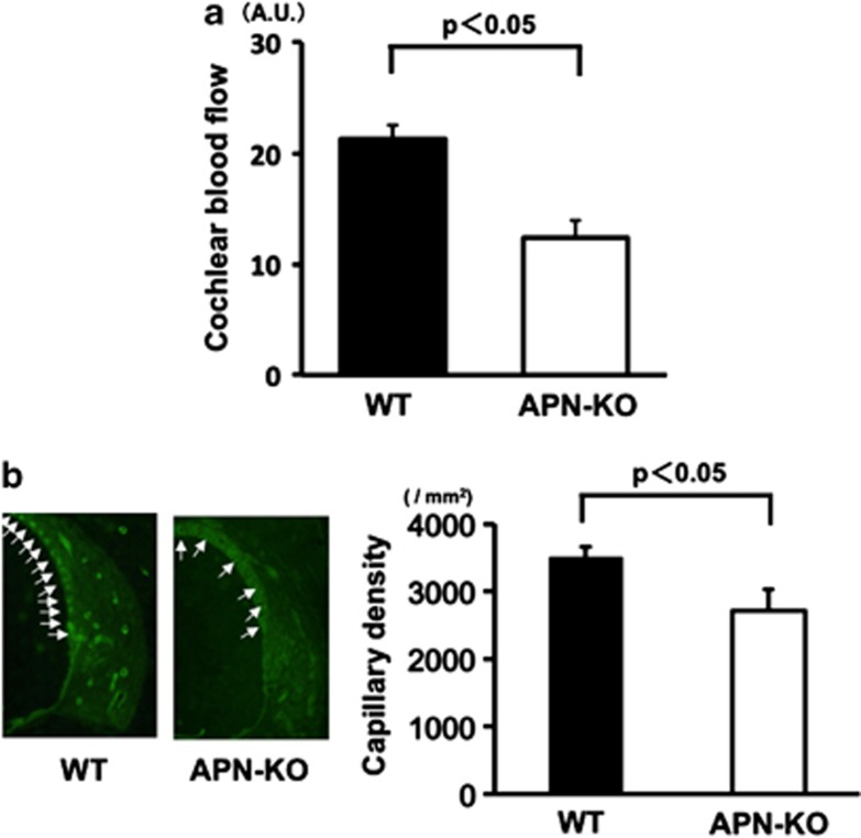
Reduced CBF and capillary density of the SV in APN-KO mice. (a) Reduced CBF in the cochlear basal turn in APN-KO mice. Quantitative analyses of CBF ratio in APN-KO and WT mice are shown. At 2 months of age, a marked reduction (48.2±4.5%) was observed in APN-KO mice as compared with WT mice (P<0.05). (b) Reduced capillary density of the SV in the cochlear basal turn in APN-KO mice. (Left panel) Fluorescence staining of the SV with an anti-CD31 monoclonal antibody (green) at 2 months of age, in WT and APN-KO mice (arrows: capillaries in the SV). (Right panel) Quantitative analysis of capillary density in APN-KO and WT mice at 2 months of age (n=4 per group). Capillary density in the SV was significantly reduced in APN-KO mice compared with WT mice at 2 months of age (P<0.05)

