Figure 3.
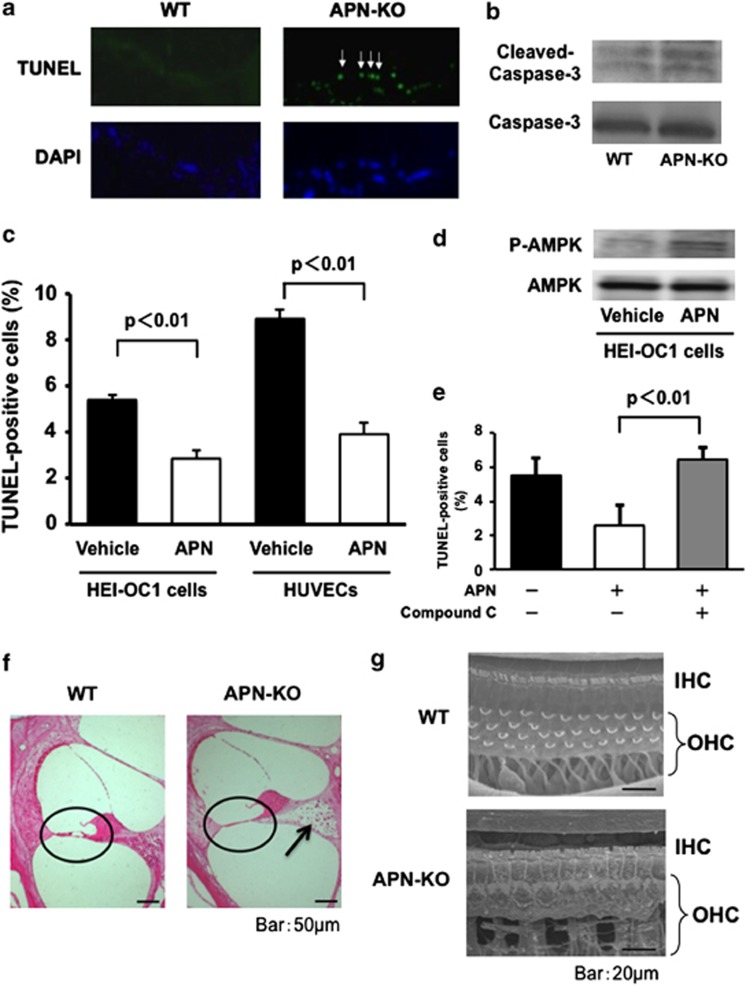
APN deficiency promotes apoptosis in the organ of Corti. (a) Representative photographs of TUNEL-positive (green) nuclei in the organs of Corti from WT and APN-KO mice at 2 months of age (arrows: hair cells) (n=4 per group). (b) The degree of cleaved caspase-3 in the organ of Corti was greater in APN-KO than WT mice at 2 months of age (n=4 per group). (c) The effect of APN on apoptosis at the cellular level. Auditory HEI-OC1 cells were subjected to hypoxia for 24 h under conditions of serum deprivation, in the presence of recombinant human APN protein or vehicle alone. Treatment with a physiological concentration of APN protein (30 μg/ml) significantly reduced the frequency of TUNEL-positive cells under hypoxic conditions (P<0.001). APN treatment also reduced the frequency of TUNEL-positive cells in HUVECs (P<0.01). (d) APN stimulates the phosphorylation of AMPK in HEI-OC1 cells. The phosphorylation of AMPK (P-AMPK) was determined by western blot analysis. Representative blots are shown from four independent experiments. (e) The effect of an AMPK inhibitor on the anti-apoptotic action of APN. HEI-OC1 cells were pretreated with compound C (10 μM) or vehicle. After treatment with compound C, HEI-OC1 cells were cultured in the presence of APN (30 μg/ml) or vehicle under conditions of hypoxia, and TUNEL-positive nuclei were quantified (n=4 per group). (f) Representative photomicrographs of the organ of Corti (encircled) in basal turns from WT and APN-KO mice at 6 months old (scale bar: 50 μm). Hair cells were lost from the basal turn of the cochlea in APN-KO mice, whereas in WT mice they were preserved. APN-KO mice also showed severe damage in the regions of spiral ganglion neurons, compared with WT mice (arrow). (g) At 6 months of age, scanning electron microscopy showed that in WT mice, three rows of OHCs and one row of IHCs were found in the organ of Corti at the basal turn of the cochlea (Figure 3d, upper panel, scale bar: 20 μm). However, the OHCs in the basal turn of the cochlea were lost in patches in APN-KO mice of the same age (Figure 3d, lower panel)

