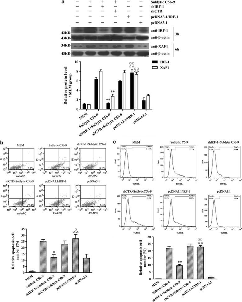
Figure 2.
The roles of IRF-1 protein expression in XAF1 gene expression and GMC apoptosis upon sublytic C5b-9 attack. GMCs were divided into six groups of (1) MEM, (2) sublytic C5b-9, (3) shIRF-1+sublytic C5b-9, (4) shCTR+sublytic C5b-9, (5) pcDNA3.1/IRF-1 and (6) pcDNA3.1. (a) The expression of IRF-1 and XAF1 in GMCs at 3 and 6 h, respectively, after sublytic C5b-9 stimulation was detected by using IB assay. (b) Both annexin V-APC and propidium iodide were used to label the cells at 6 h after sublytic C5b-9 stimulation. The number of annexin V-positive GMCs was found by using flow cytometry analysis. (c) TUNEL staining (TMR-labeled) was used to label the apoptotic cells in different groups at 6 h after sublytic C5b-9 stimulation. Flow cytometry analysis was performed to detect the numbers of TUNEL-positive GMCs (n=3 in each group). The data are from one experiment, representative of three independent experiments. Results were represented as means±S.E. (n=3 in each group). Representative photographs were manifested. *P<0.05, **P<0.01 versus sublytic C5b-9 group and shCTR+ sublytic C5b-9 group; ΔΔP<0.01 versus MEM group; □P<0.05, □□P<0.01 versus pcDNA3.1 group

