Figure 6.
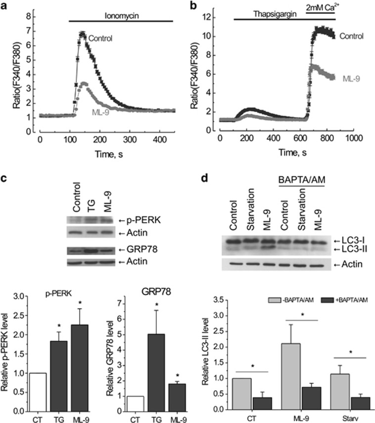
ML-9 modulates Ca2+ homeostasis and stimulates autophagy in a calcium-dependent manner. (a) LNCaP cells were pretreated with full medium or 30 μM ML-9-containing medium for 6 h, loaded with Fura2/AM probe and subjected to calcium imaging experiment. 2 μM ionomycin-induced transients in calcium-free extracellular medium were analyzed. (b) ML-9 reduces both calcium content of intracellular calcium stores and SOCE in LNCaP cells. LNCaP cells were treated as in (a). 2 μM TG-induced transients in calcium-free extracellular medium as well as SOCE were analyzed. (c) ML-9 induces ER-stress. LNCaP cells were treated with full medium, 1 μM TG-containing medium or 50 μM ML-9-containing medium for 24 h. The levels of p-PERK and GRP78 were analyzed. Densitometric quantitation for normalized p-PERK and GRP78 relative to Actin is shown. Values represent means±S.E.M. n=3. (d) Ca2+ is required for ML-9-induced autophagy. LNCaP cells were treated with full, serum-starved, 30 μM ML-9-containing full media in the absence or presence of 20 μM BAPTA/AM for 3 h. Densitometric quantitation for normalized LC3-II relative to Actin is shown. Values represent means±S.E.M. n=3

