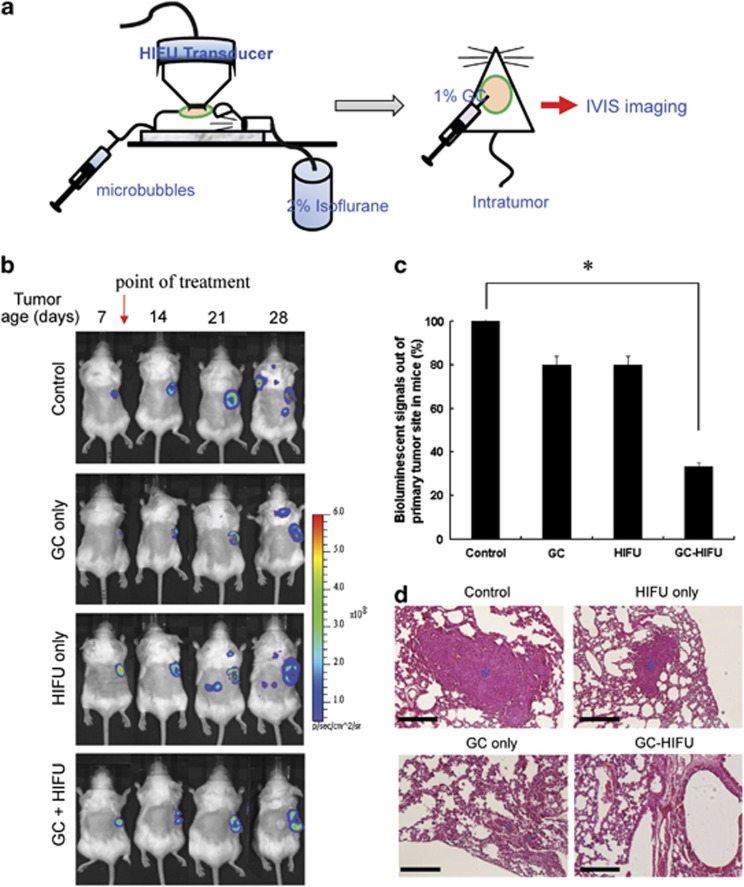
Figure 3.
Suppression of lung metastasis of 4T1_PB3R cells in female Balb/c mice by GC-HIFU treatment. (a) Schematics of GC-HIFU treatment. (b) Progression of 4T1_PB3R tumors in mice under different treatments, determined by IVIS imaging system (N=6 for each experimental group). (c) Bioluminescent signals from non-primary tumor sites among different experimental groups, in comparison with that of untreated control group, according to the IVIS data. *P<0.05. (d) Tumor metastasis in lung sections detected using hematoxylin and eosin staining (marked with asterisks). The scale bar of each picture is 200 μm

