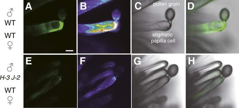
Figure 3.
Detection of ROS on Stigmatic Papilla Cells.
Detection of ROS is shown on stigmatic papilla cells with Oxyburst Green 20 min after pollination.
(A) to (D) Intense fluorescence of Oxyburst Green around a wild-type pollen tube growing within the stigmatic papilla cell wall.
(E) to (H) Faint fluorescence of Oxyburst Green around the rbohH-3 rbohJ-2 double mutant pollen tube growing within the stigmatic papilla cell wall.
(A) and (E) show fluorescence images; (B) and (F) show pseudocolor fluorescence images; (C) and (G) show bright-field images; and (D) and (H) show overlay images. Bar = 10 μm.

