Figure 4.
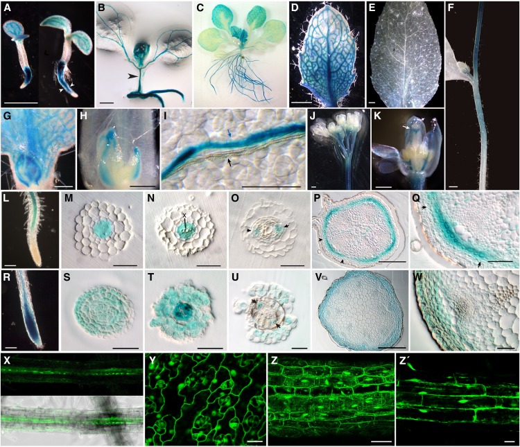
D14 Promoter Activity and D14 Protein Distribution during Arabidopsis Development.
GUS histochemical activity of Arabidopsis D14pro:GUS ([A] to [Q]) and D14pro:D14:GUS ([R] to [W]) transgenic plants.
(A) Five-day-old transgenic seedlings. The plant on the right, more advanced in development, shows expression in the root more restricted to the vascular cylinder (arrowheads) than that of the less developmentally advanced (left).
(B) Ten-day-old seedling with GUS activity in the vascular tissue of the hypocotyl (arrowhead).
(C) Eighteen-day-old vegetative rosette.
(D) Young rosette leaf from plant in (C).
(E) Mature cauline leaf from 30-day-old plant.
(F) Stem of the main inflorescence showing a gradient of GUS activity with a maximum near the apex.
(G) Bud in the axil of a young rosette leaf.
(H) Bud in the axil of a mature rosette leaf.
(I) Detail of a rosette leaf surface. Note the separation between the xylem (white) bundle (black arrow) and the phloem (blue) bundle expressing GUS (blue arrow).
(J) Main inflorescence. GUS accumulates in the apical-most stem region and in flower pedicels.
(K) Close-up of a developing flower. Signal in the style is indicated (white arrow).
(L) Root tip.
(M) to (O) The 3-μm transverse plastic-embedded sections of root similar to that in (L).
(M) Distal section showing GUS staining in procambium cells.
(N) GUS is excluded from xylem cells (X).
(O) More proximal section showing promoter activity in phloem cells (arrows).
(P) Transverse plastic-embedded section of a stem internode of the primary inflorescence.
(Q) Close-up of a section similar to that shown in (M), with cortex cells but not epidermis cells expressing GUS. Notice the stronger signal in the vascular bundle sector flanked by the arrows in (P) and (Q).
(R) Root tip similar to that in (L). D14:GUS is present in the root tip.
(S) to (U) The 3-μm transverse plastic-embedded sections of root tips. (S) shows the meristematic zone, and (T) and (U) are sections similar to those in (N) and (O). GUS signal is widespread in (S) and (T) and accumulates in the epidermis, cortex, and phloem (arrowheads) in (U).
(V) and (W) Stem transverse plastic-embedded sections comparable to those in (P) to (Q). GUS is detectable throughout the cortex, epidermis, and phloem (arrowheads).
(X) GFP fluorescence image (top) and fluorescence merged with bright-field image (bottom) of a transgenic D14pro:D14:GFP root.
(Y) to (Z’) Leaf (Y), hypocotyl (Z), and root (Z’) cells of CaMV35Spro:D14:GFP transgenic plants. GFP is detected in nucleus and cytoplasm.
Bars = 1 mm in (A), (D) to (F), and (K), 500 μm in (L), 200 μm in (B), (M), and (P), 100 μm in (G) and (N), 50 μm in (H) to (J), (Q), and (Z), 15 μm in (Y) and (Z’).

