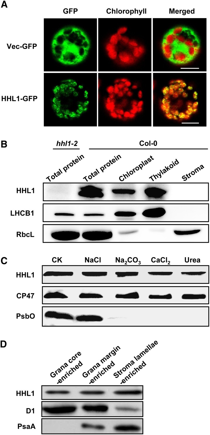
Figure 4.
Subcellular Localization and Immunolocalization of HHL1 Protein.
(A) Localization of HHL1 protein within the chloroplast by GFP assay. The fluorescence of HHL1-GFP specifically matched with that of chlorophyll autofluorescence, confirming chloroplast targeting of HHL1 exclusively. HHL1-GFP, HHL1-GFP fusion; Vec-GFP, control with empty vector. Bars = 10 μm.
(B) HHL1 localizes to the thylakoid membrane fractions. Intact chloroplasts were isolated from wild-type (Col-0) leaves and then separated into thylakoid membrane and stroma fractions. Polyclonal antibodies were used against the integral membrane protein LHCB1, the abundant stroma protein ribulose biphosphate carboxylase large subunit (RbcL) and HHL1. Total proteins extracted from the wild type and hhl1-2 were used to confirm the specificity of the anti-HHL1 antibody.
(C) Immunolocalization of HHL1. The wild-type thylakoid membranes were sonicated in the presence of 1 M NaCl, 200 mM Na2CO3, 1 M CaCl2, and 6 M urea for 30 min at 4°C. PsbO (the 33-kD luminal protein of PSII) and CP47 (the PSII core protein) were used as markers. Membranes that had not been subjected to any salt treatment were used as controls (CK).
(D) Analysis of HHL1 protein in grana core-, grana margin-, and stroma lamellae-enriched thylakoid. Thylakoids were solubilized with digitonin and subfractionated by differential ultracentrifugation into grana core-, grana margin-, and stroma lamellae-enriched thylakoid and subjected to immunoblot analysis. The lanes on each gel were loaded on an equal chlorophyll basis. All experiments were repeated three times with similar results.

