Figure 6.
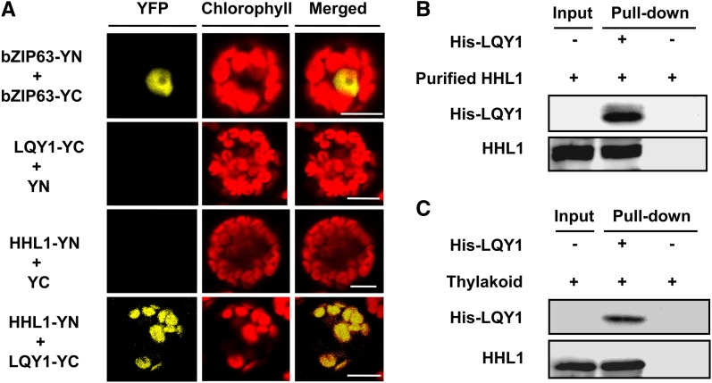
BiFC Visualization and Pull-Down Experiments Show Interaction between HHL1 and LQY1 in Vivo and in Vitro.
(A) BiFC analysis of Arabidopsis protoplasts showing the interaction between HHL1 and LQY1 in vivo. HHL1 fused with the N terminus of YFP (YN) and LQY1 fused with the C terminus of YFP (YC) were cotransfected into protoplasts and visualized using confocal microscopy. As a positive control, both bZIP663 fused with YN and bZIP663 fused with YC were cotransfected into protoplasts. As negative controls, HHL1 fused with YN and empty vector YC as well as LQY1 fused with YC and empty vector YN were cotransfected into protoplasts. Bars = 10 μm.
(B) and (C) Pull-down assays showing HHL1 interaction with LQY1 in vitro. His-LQY1 bound to CNBr-activated resin was incubated with recombinant HHL1 expressed in E. coli (B) and DM-solubilized thylakoid membranes (C). Bound proteins were eluted, separated by SDS-PAGE, and subjected to immunoblot analysis with His-tag and HHL1 antibodies. All experiments were repeated at least two times with similar results.

