Figure 7.
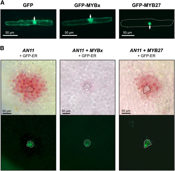
Localization and Intercellular Movement of MYB Repressors and AN11.
(A) Biolistic transformation of onion bulb scale epidermis with 35Spro:GFP, 35Spro:GFP-MYBx, or 35Spro:GFP-MYB27 viewed with blue light on a dissecting microscope; arrows indicate the nucleus and the cell margins are indicated (dotted boundary) for GFP-MYB27.
(B) Biolistic transformation of petunia W134 (an11) petals with 35Spro:AN11, 35Spro:AN11 + 35Spro:MYBx, or 35Spro:AN11 + 35Spro:MYB27. A 35Spro:GFP-ER construct was included in all transformation as an internal control to label the transformed cells; GFP was viewed with blue light. The transformed cell is indicated (dotted boundary).

