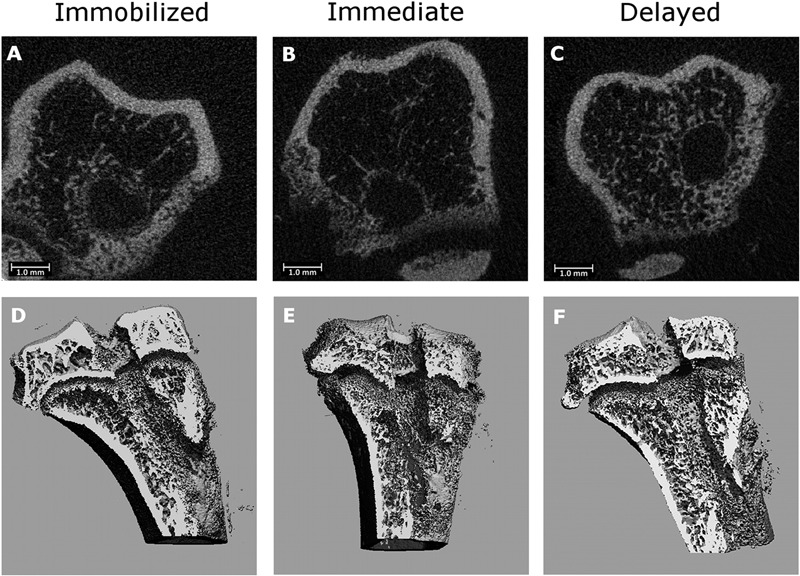
Fig. 2.

Representative axial micro-CT images of the tibial tunnel after immobilization (Fig. 2-A), immediate high-strain loading (Fig. 2-B), and delayed high-strain loading (Fig. 2-C). Three-dimensional reconstructions of the tibial tunnel after immobilization (Fig. 2-D), immediate high-strain loading (Fig. 2-E), and delayed high-strain loading (Fig. 2-F). There is greater bone formation in the immobilized and delayed-loading groups than in the immediate-loading group.
