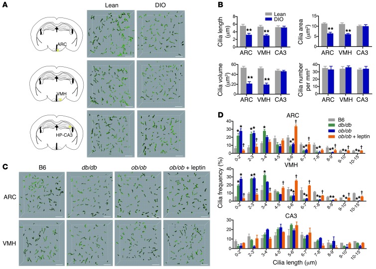
Figure 1. Reduced cilia length in the hypothalamus of adult obese mice.
(A and B) 3D-reconstructed cilia images (A) and analysis of cilia parameters (B) in ARC, VMH, and hippocampal region CA3 of 20-week-old lean or DIO mice (n = 5 per group). (C and D) AC3-immunoreactive cilia images (C) and cilia distribution (D) in ARC, VMH, and hippocampal region CA3 in 12-week-old lean C57BL/6 (B6) or obese (ob/ob and db/db) mice and in ob/ob mice treated with leptin (10 μg/d) for 7 days (n = 3–4 per group). Data represent mean ± SEM. *P < 0.05, **P < 0.005 vs. lean control; †P < 0.05 vs. untreated ob/ob. Scale bars: 15 μm.

