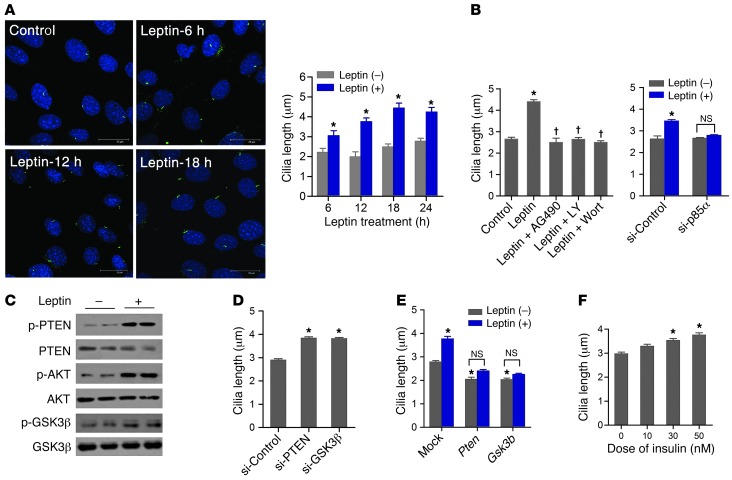
Figure 2. Leptin stimulates cilia assembly in hypothalamic neuron cells.
(A) Acetylated α-tubulin immunocytochemistry and cilia length measurement in N1 hypothalamic neuron cells treated or not with 100 nM leptin for the indicated times. Scale bars: 25 μm. (B) Changes in cilia length in N1 cells treated with leptin (100 nM for 18 hours) and/or AG490 (1 μM), LY294002 (1 nM), or wortmannin (10 nM), or in cells transfected with control siRNA (si-Control) or Pik3r1 siRNA (si-p85α). (C) Leptin treatment (100 nM for 30 minutes) of N1 cells caused serial phosphorylation of PTEN, AKT, and GSK3β in N1 cells. (D) siRNA-mediated knockdown of PTEN and GSK3β increased cilia length in N1 cells. (E) Effect of leptin treatment (100 nM for 18 hours) on cilia length in N1 cells overexpressing Pten and Gsk3b. (F) Effect of insulin treatment (18 hours) on cilia length in N1 cells. Data are mean ± SEM. *P < 0.05 vs. control; †P < 0.01 vs. leptin alone.

