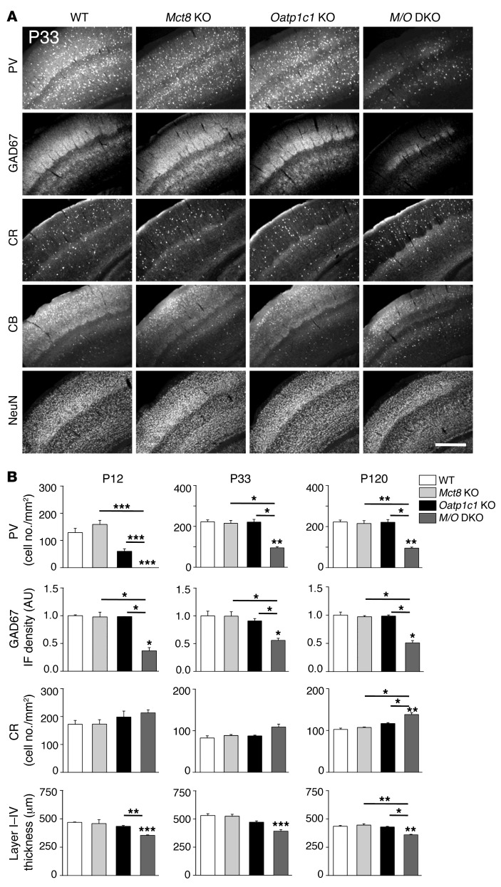
Figure 7. Histological analysis of GABAergic neurons in the somatosensory cortex.
Coronal forebrain vibratome sections from perfusion-fixed male mice (n = 3 per genotype and time point) were immunostained with antibodies recognizing the calcium-binding proteins PV, CB, and CR; the neuronal transcription factor NeuN; and the GABA-producing enzyme GAD67. (A) Representative views demonstrating visibly reduced P33, PV, and GAD67 immunoreactivity in the somatosensory cortex of Mct8/Oatp1c1 DKO mice. Scale bar: 250 μm. (B) This optical impression was confirmed by measuring GAD67 integrated fluorescence signal density and counting PV-positive cells using ImageJ. Quantification also revealed a slight increase in the number of CR-positive neurons in Mct8/Oatp1c1 DKO mice. Thickness of the cortical layers was determined in NeuN-stained brain sections and disclosed a thinner layer I–IV in Mct8/Oatp1c1 DKO mice. Overall, these data pointed to pronounced alterations in the cortical GABAergic system in Mct8/Oatp1c1 DKO animals. *P < 0.05, **P < 0.01, ***P < 0.001 vs. WT, or as otherwise indicated (brackets).

