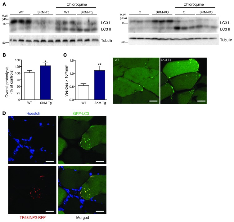
Figure 3. TP53INP2 increases basal autophagy in skeletal muscle by inducing the formation of autophagosomes.
(A) Western blot analysis of LC3I and LC3II content in total homogenates of gastrocnemius muscles from WT and SKM-Tg mice or from control and SKM-KO mice. Representative images are shown. Mice were treated with chloroquine as indicated. (B) Overall proteolysis assessed as the rate of tyrosine released to the media in incubated extensor digitorum longus muscles (n = 14). (C) Autophagosomes were quantified by counting EGFP-LC3–positive dots normalized for cross-sectional area from tibialis anterior muscles transfected with EGFP-LC3 from WT and SKM-Tg mice fasted for 16 hours (n = 5). Representative images of transverse sections from adult tibialis anterior muscles transfected with EGFP-LC3 of WT and SKM-Tg mice are shown. Scale bar: 15 μm. (D) Confocal images of tibialis anterior muscle transverse sections showing colocalization between TP53INP2 and LC3. Adult muscles were transfected with TP53INP2-RFP and EGFP-LC3. TP53INP2 is stained in red, LC3 is stained in green, and nuclei are stained in blue (Hoechst33342 staining). Scale bar: 30 μm. Data represent mean ± SEM. *P < 0.05, **P < 0.01 vs. control values.

