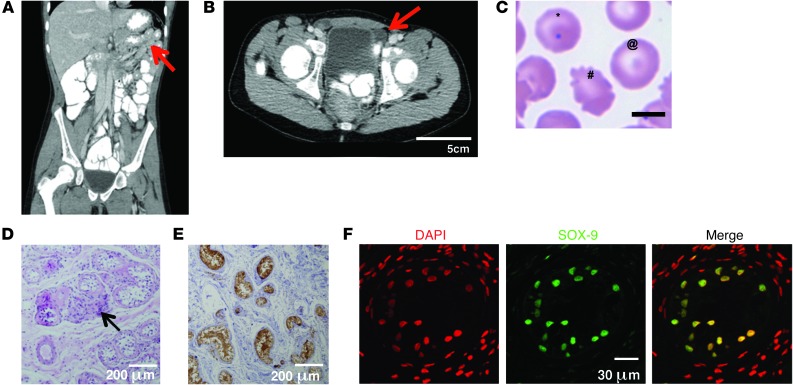
Figure 1. Phenotypic features of the patient.
(A) CT scan of the patient’s abdomen, revealing asplenia (arrow) and lack of uterus. (B) CT scan of the inguinal region, revealing inguinal testes (arrow). (C) Patient’s peripheral blood smear, with Howell-Jolly bodies (*), target cells (@), and poikilocytosis (#) indicative of asplenia. (D) H&E staining showed paucity of Leydig cells (arrow), lack of germinal cells, and abundance of Sertoli cells. (E) Paraffin-embedded sections of the patient’s testes immunostained with anti-inhibin antibody (brown). (F) Immunofluorescent staining of the proband’s testis with anti–SOX-9 antibody (green) and general nuclear staining with DAPI (red). Positive SOX-9 staining in Sertoli cells was similar in strength in the patient’s testes and in the control testes, which had a higher total number of cells, consisting mostly of germinal and pregerminal cells (not shown). Scale bars: 5 μm (C); 200 μm (D and E); 30 μm (F).

