
Figure 1.
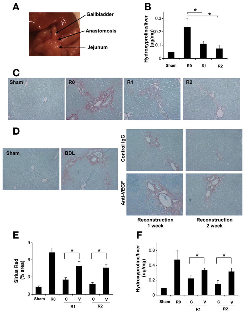
Anti-VEGF antibody disrupts fibrosis resolution. C57BL/6 mice were subjected to BDL for 2 weeks. CJ was performed to reconstruct biliary flow and induce fibrosis resolution. Reconstructed anatomy 2 weeks after CJ is shown (A). Hydroxyproline content (B) and Sirius Red staining (200×) (C) demonstrated almost complete fibrosis resolution two weeks after CJ. Mice received VEGF-neutralizing antibody or control antibody (IP ×2/week for 1 or 2 weeks), after 2 weeks of BDL followed by CJ. One week and 2 weeks after CJ, livers were harvested and subjected to analysis. Fibrosis resolution was attenuated by anti-VEGF assessed by Sirius Red staining (D), Sirius Red quantification (E), and hydroxyproline content (F). (R0: BDL 2 week without CJ; R1: BDL 2 weeks plus CJ for 1 more week; R2: BDL 2 weeks plus CJ for 2 more weeks; C: control IgG; V: anti-VEGF antibody; n = 10; *P < .05).