
Figure 2.
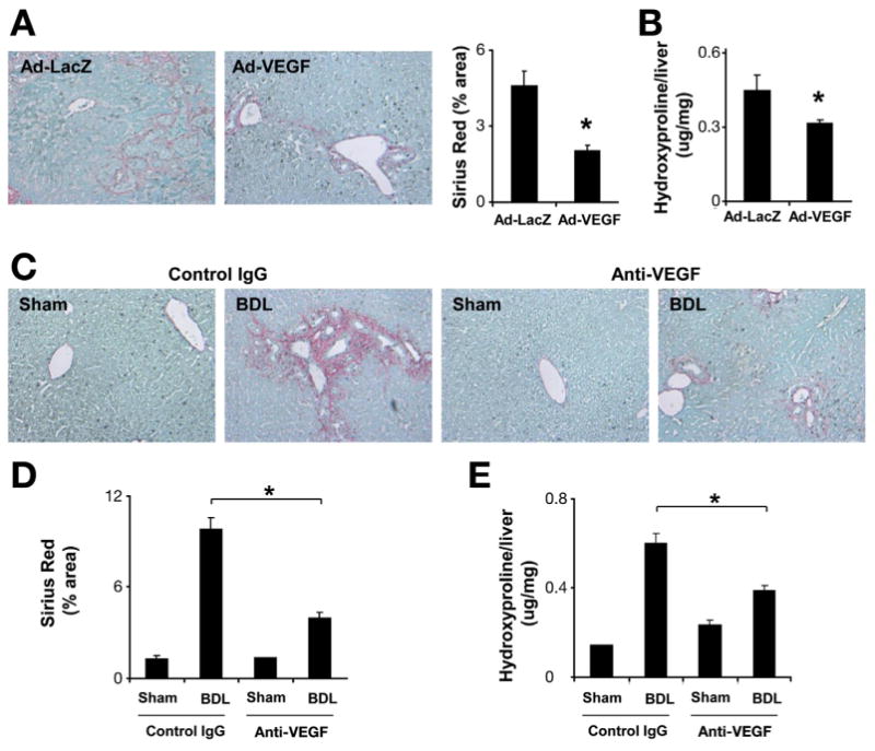
VEGF overexpression promotes fibrosis resolution. C57BL/6 mice were subjected to BDL for 2 weeks followed by CJ. One day after CJ, adenovirus-expressing mouse VEGF or LacZ (single dose 0.8 × 10⁹ PFU/kg) was injected through tail vein injection. All animals were sacrificed 1 week after CJ. Fibrosis was assessed by Sirius Red staining (200×) (A) and hydroxyproline assay (B). VEGF overexpression was associated with enhanced fibrosis resolution (n = 7;*P < .05). C57BL/6 mice were subjected to BDL. One day after BDL, C57BL/6 mice received VEGF-neutralizing antibody or control antibody (IP ×2/week for 2 weeks). Two weeks after BDL, animals were sacrificed. Sirius Red staining (C) and quantification (D) showed decreased liver fibrosis after neutralizing anti-VEGF antibody treatment. Hydroxyproline quantification is shown as well (E) (n = 8; *P < .05).