
Figure 5.
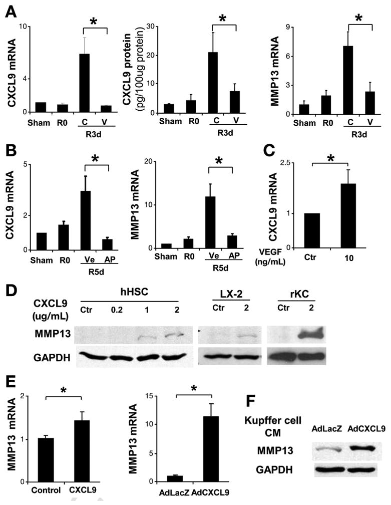
VEGF-neutralizing antibody abrogates CXCL9 and MMP13 production during fibrosis resolution. After BDL + CJ, C57BL/6 mice received 1 dose of VEGF-neutralizing antibody or control antibody. Three days later, mice were sacrificed and total liver mRNA and protein was isolated. CXCL9 mRNA and protein levels were significantly elevated during fibrosis resolution and this was abrogated by anti-VEGF antibody (A). MMP13 mRNA levels were elevated during fibrosis resolution and this was attenuated by anti-VEGF antibody (A). After CJ + BDL, MAFIA mice were treated with vehicle or AP20187 daily for additional 5 days. Total liver mRNA was subjected to real-time polymerase chain reaction for CXCL9 and MMP13. CXCL9 and MMP13 (B) elevation during fibrosis resolution was attenuated after Kupffer cell depletion with AP20187 in MAFIA mice. Fresh isolated Kupffer cells were treated with VEGF (10 ng/mL) or control for 3 hours. CXCL9 mRNA levels were elevated compared with control (C). Human HSC (hHSC) were treated with human recombinant CXCL9 for 48 hours. Total cell lysates were subjected to Western blot analysis. MMP13 increases in response to increasing concentrations of CXCL9 as shown (D). LX2 cell line was tested under same conditions with similar results (D). Freshly isolated rat Kupffer cells were treated with recombinant mouse CXCL9 for 48 hours. MMP13 production was evaluated by Western blot analysis (D). Human primary hepatic stellate cells (hHSC) were treated with recombinant CXCL9 for 48 hours after overnight serum starvation in 0.5% fetal bovine serum Dulbecco's modified Eagle medium. MMP13 mRNA levels were elevated in response to CXCL9 (E). hHSC were transfected with control Ad-LacZ or Ad-CXCL9 (multiplicity of infection = 40). After 48 hours, MMP13 mRNA expression was increased in response to Ad-CXCL9 (E). Culture-activated rat HSC was treated with Kupffer cell condition medium infected with Ad-LacZ and Ad-CXCL9. Western blot for CXCL9 was shown in (F). R0: BDL 2 weeks without CJ; R3d: BDL 2 weeks plus CJ for 3 days; R5d: BDL 2 weeks plus CJ for 5 days; C, control IgG; V, anti-VEGF antibody; Ve, vehicle; AP, AP 20187; n = ; *P <.05).