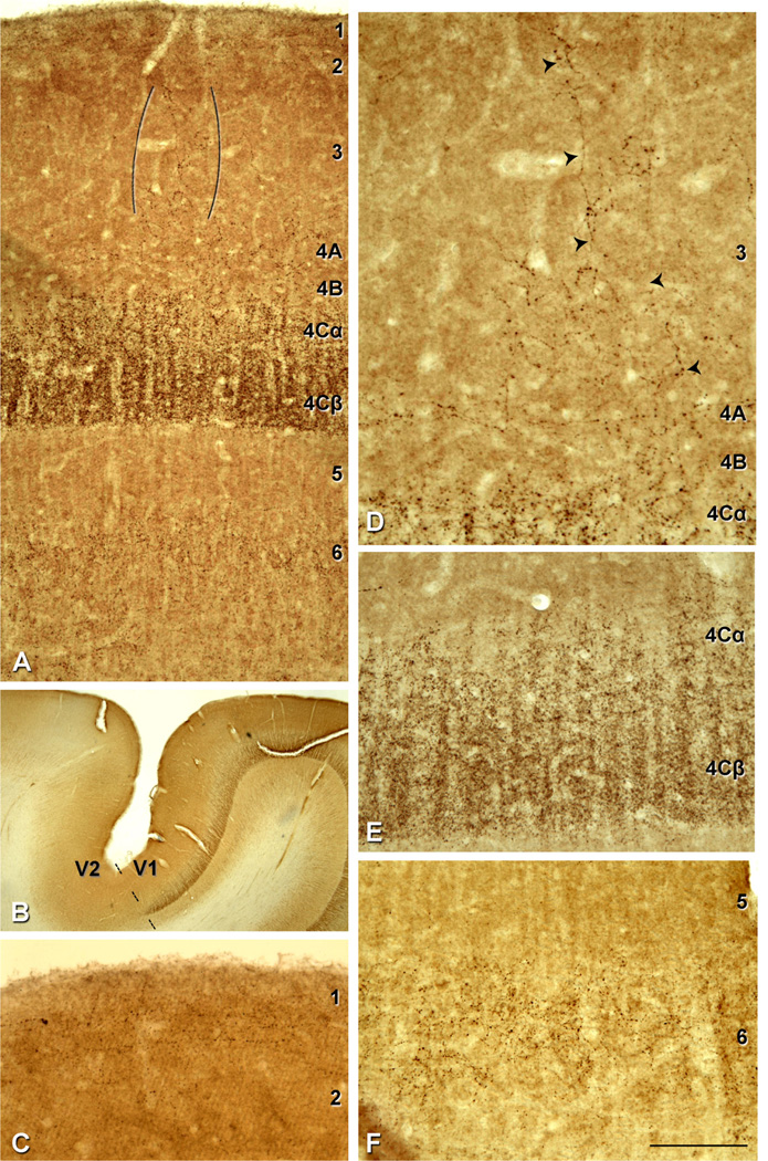
Figure 2.
VGluT2-ir pattern in human V1. A: Immunostaining in layers 1, 2/3, 4A, 4Cα, 4Cβ and 6. VGluT2-ir puncta in layer 3 (indicated between brackets). B: V1/V2 border (dashed line) stained for VGluT2. There is a very clear laminar pattern of VGluT2-ir in V1 that ends abruptly at the V2 border. C: VGluT2-ir puncta in layer 1 prominently found in the upper half of the layer. D: Higher magnification of VGluT2-ir puncta fibrous pattern in layer 3 (indicated by the arrowheads). E: Higher magnification of layers 4Cα and 4Cβ; due to the high density of VGluT2-ir puncta in these layers cortical minicolumns are clearly observed. F: Higher magnification of VGluT2-ir puncta in layer 6. Scale bar in F = 200 µm for A; 2,150 µm for B; 145 µm for C,E,F; 110 µm for D.

