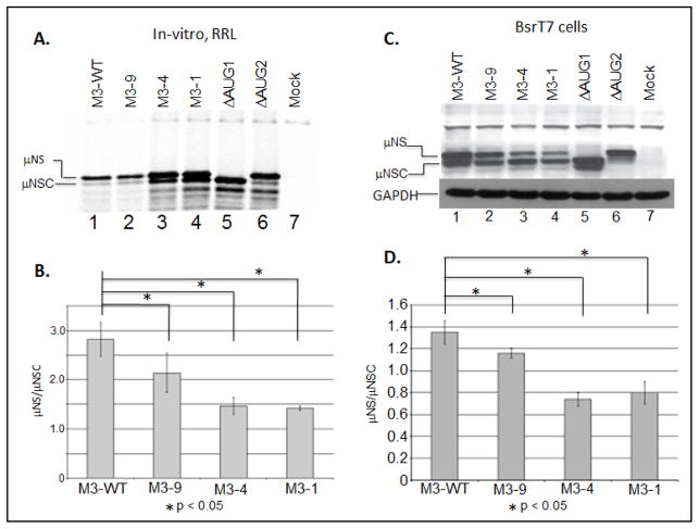
Figure 2. The MRV M3 mRNA 5′ UTR is not necessary for translation initiation but does influence start site selection.
A) In-vitro RRL translation reactions were programmed with 2 μg of M3-WT, M3-9, M3-4, or M3-1 mRNAs. Reactions were incubated with [35S]methionine at 30°C for 90 minutes. Reactions were stopped by addition of 2X Laemmli sample buffer and labeled proteins were separated by 8% SDS-PAGE. Labeled proteins were visualized by phosphorimaging. B) Radiolabeled μNS and μNSC proteins were quantified using ImageQuant 7.0 software. μNS/μNSC ratios were determined for each mRNA construct. μNS/μNSC ratios are presented as the arithmetic mean ± SD of three independent assays in which samples were analyzed in triplicate. Significance was determined by Student’s t-test where P<0.05 was considered significant. C) In-vitro transcribed M3-WT and mutant construct mRNAs were transfected into BsrT7 cells. At 24h p.t. cells were lysed and μNS, μNSC and GAPDH proteins were detected by Immunoblotting. D) μNS and μNSC protein concentrations were determined by densitometry, and μNS/μNSC ratios were calculated and plotted. μNS/μNSC ratios are presented as the arithmetic mean ± SD of three independent assays with different preparations of RNA in which samples were analyzed in triplicate. Significance was determined by Student’s test where P<0.05 was considered significant.

