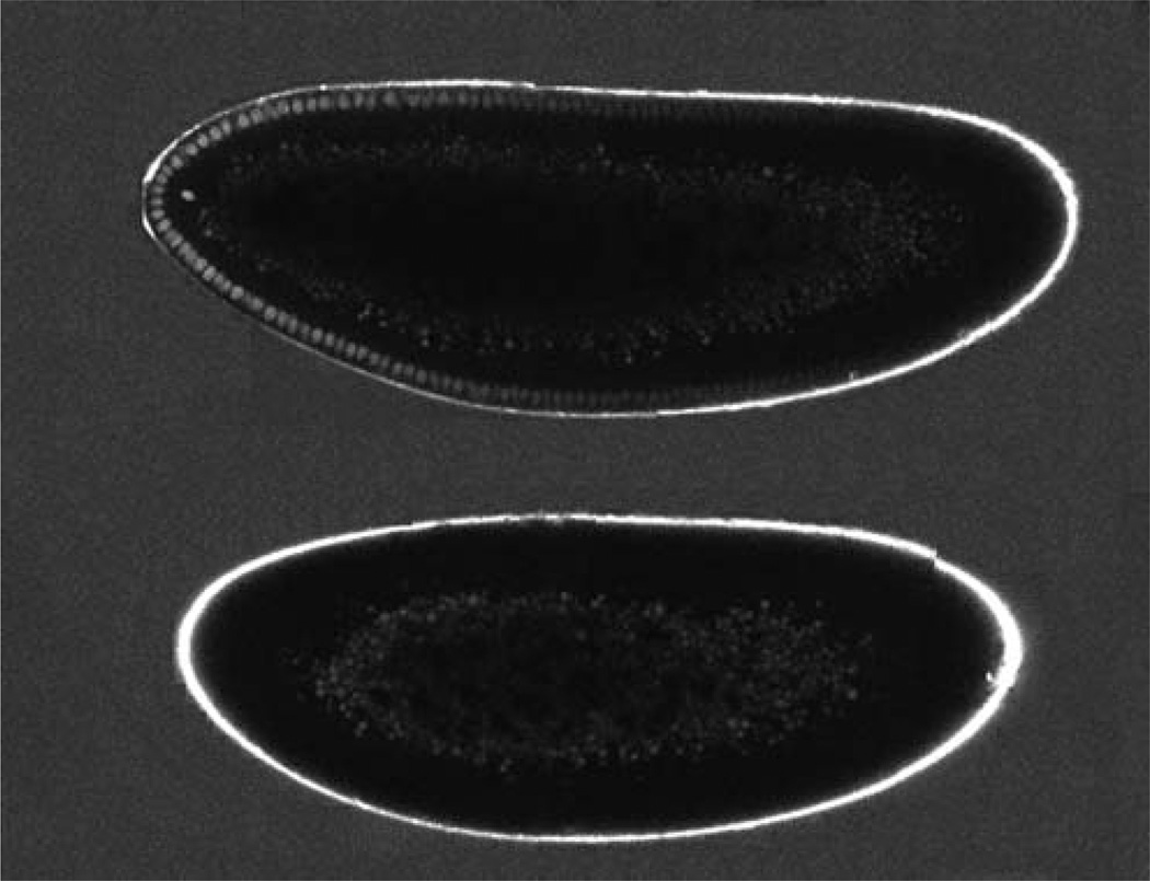
FIGURE 3.
Absolute concentration measurements. An embryo expressing a Bcd–GFP fusion protein (top) and a wild-type embryo (bottom) immersed in a solution of 34 ± 3 nM GFP. Both embryo images were taken during the same imaging session; each embryo was imaged in three pieces, which were reassembled by software. The two resulting embryo images were joined for display. (Note that the difference in embryo size reflects naturally varying egg sizes in the wild-type population.)

