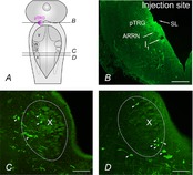
Figure 6. Retrogradely labelled neurons in the vagal motoneuron region.

A, schematic illustration of a dorsal view of the lamprey mesencephalon–rhombencephalon showing the levels of the coronal sections illustrated in B–D (continuous lines) and the location of the pTRG (pink area). B, photomicrograph of a transverse section of the isthmic region showing the site of neurobiotin injection into the pTRG (green signal). C and D, photomicrographs of transverse sections, 100 μm apart, of the vagal motoneuron region (dotted line) showing retrogradely labelled neurons (green signal) after an injection of neurobiotin into the pTRG. Arrowheads point to some retrogradely labelled neurons. ARRN, anterior rhombencephalic reticular nucleus; I1, isthmic Müller cell; pTRG, paratrigeminal respiratory group; SL, sulcus limitans of His; V, trigeminal motor nucleus; VII, facial motor nucleus; IX, glossopharyngeal motor nucleus; X, vagal motor nucleus. Scale bars, 200 μm in B, 100 μm in C and D.
