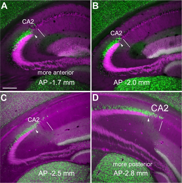
Figure 5.
PCP4 staining differs along the anterior–posterior axis across coronal sections of adult mouse hippocampus. A–D: The overlaid images (PCP4, green and mossy fiber tract, magenta) of the coronal sections at different anterior–posterior (AP) coordinates show more widespread PCP4 immunolabeling at the posterior sections. The PCP4 staining was done on the sections of Calb2-Cre:tdTomato mice, in which dentate granule cells express red fluorescent proteins in their mossy fibers. The arrowhead indicates the end of the mossy fiber tract, and the thin white line indicates the border between CA2 and CA1. Scale bar = 200 μm in A (applies to A–D).

