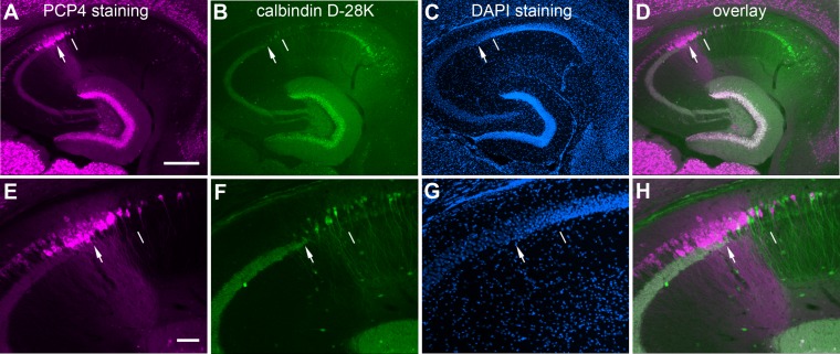
Figure 6.
Double immunolabeling of mouse hippocampal sections against PCP4 and calbindin D-28K (CB) further helps to determine the distal CA3/CA2 border. A–H: The images of PCP4 immunostaining (A,E), CB staining (B,F), DAPI staining (C,G), and PCP4/CB staining overlays (D,H) of one example hippocampal section, taken under lower (A–D) versus higher power (E–H) objectives. The arrowhead indicates the end of the mossy fiber tract, and the thin white line indicates the border between CA2 and CA1. Scale bar = 200 μm in A (applies to A–D); 50 μm in E (applies to E–H).

