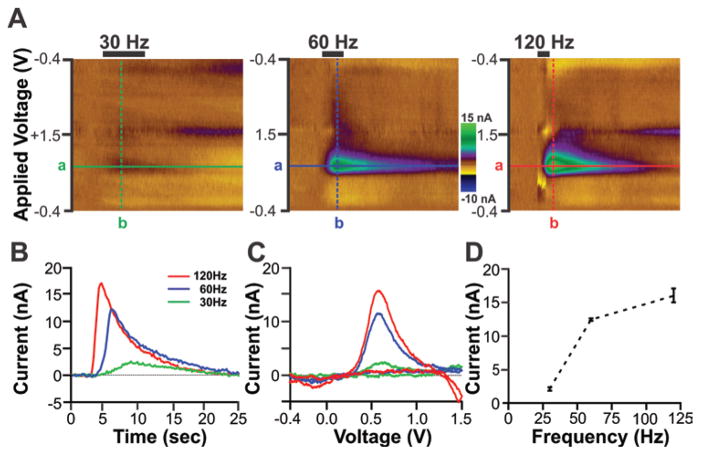
Fig. 5.
In vivo functionality tests of the integrated MINCS-WINCS devices showing frequency-dependent striatal dopamine release evoked by MFB electrical stimulation in anesthetized rats. A: Color plots obtained in response to electrical stimulation delivered at 30 Hz (left), 60 Hz (center), and 120 Hz (right). The number of pulses (120), stimulation intensity (200 μA), and pulse duration (2 msec) for the biphasic stimulation train were the same for each test frequency. The FSCV triangle waveform was applied from −0.4 V to +1.5 V with dopamine oxidation peak currents readily apparent at +0.6 V. B: Time course of changes in dopamine oxidation current in response to 30-, 60-, and 120-Hz electrical stimulation. These changes in dopamine oxidation currents were detected at the applied voltage (+0.6 V) shown in each color plot (line a). C: Voltammograms of dopamine oxidation and reduction after subtraction of prestimulation background current. These voltammograms were extracted at the time corresponding to maximal increases in dopamine oxidation current (line b). D: Stimulation frequency–dependent striatal dopamine release showing an exponential increase in mean ± SEM peak dopamine oxidation current as a function of increasing stimulation frequency. Note that the data shown in panels A–C are from a representative animal and data are the mean ± SEM of 3 stimulations/frequency in the same animal.

