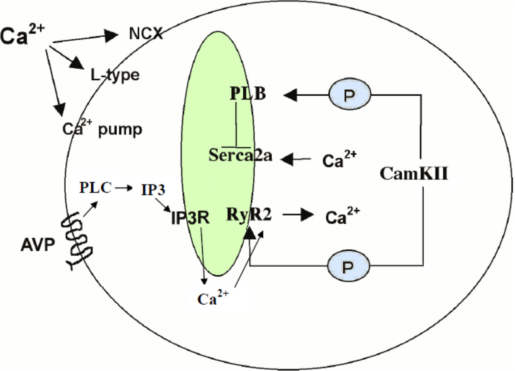
Figure 1. Proteins in the cardiomyocyte Ca2+ Signaling Pathway.
A typical contraction in a mammalian cardiomyocyte is initiated by Ca2+ entry into the cytosol via L-type Ca2+ channels on the sarcolemma. This Ca2+ influx can directly cause release of Ca2+ from the sarcoplasmic reticulum via the ryanodine receptor (RyR2) channel on the sarcoplasmic reticulum (SR) resulting in an increased [Ca2+]i. The high levels of [Ca2+]i, in turn, trigger the myofilaments to contract and result in activation of CamKIIγ which can phosphorylates phospholamban to release its inhibition from Serca2a. Serca2a then pumps Ca2+ from the cytosol into the SR for the next cardiac cycle. Ca2+ signaling can also be initiated by vasopressin through the G-protein coupled AVP receptor to activate the IP3 pathway. Local production of IP3 results in release of Ca2+ form the SR via the IP3 receptor (IP3R) Ca2+ channel. The removal of cytosolic Ca2+ is also through Serca2a.

