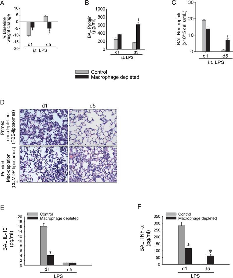
Fig. 6. IL-10 producing alveolar macrophages are critical for immunological priming.
Primed mice treated with PBS-liposomes (control) and primed mice treated with CL2-MDP liposomes (macrophage depleted) were assessed for body weight relative to baseline (A), BAL total protein (B), BAL neutrophils (C) or histological damage (D) by H&E staining (x100 magnification) at days 1 or 5 after i.t. LPS injury. BAL IL-10 (E) and TNF-α from primed control and primed macrophage-depleted were measured at days 1 or 5 after i.t. LPS. Values expressed as mean ± SEM; *one-way ANOVA (A-C) or paired t-test (E-F) against other groups at same time point, p<0.05, (n=4-6 animals per group per time point)

