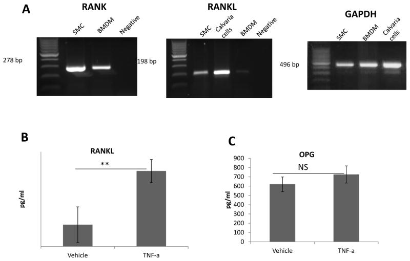
Figure 2. RANK, RANKL and OPG expression.
(A) RT-PCR with RNA isolated from SMCs, BMDMs and positive control calvaria cells. RANK was highly expressed in both SMCs and BMDMs. RANKL was also highly expressed in SMCs but not in BMDMs. Amplification of GAPDH was used to determine the quality of the RNA. (B) ELISA for RANKL and OPG in conditioned medium collected from SMCs. Cells were treated with vehicle or TNF-α for 48 hours. Very low RANKL protein was detected, **<0.05. In contrast, very high levels of OPG were present.

