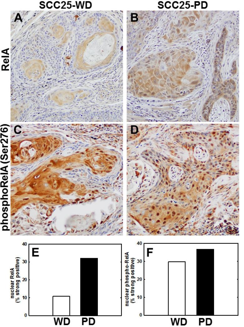
Figure 4. Nuclear staining for RelA and phospho-RelA(Ser276) is enhanced in SCC25-PD tumors relative to SCC25-WD tumors.
(A,B) Immunohistochemical analysis of tumor section from orthotopic (tongue) mouse xenograft model initiated with (A) SCC25-WD cells or (B) SCC25-PD cells stained with antibodies directed against RelA. (C,D) Immunohistochemical analysis of tumor section from orthotopic murine xenograft initiated with (C) SCC25-WD cells and (D) SCC25-PD cells stained with antibodies directed against phospho-RelA(Ser276). Magnification – 200X. (E) Quantitation of nuclear RelA staining. (F) Quantitation of nuclear phospho-RelA(Ser276) staining. (Detailed quantitation results are shown in Supplementary Table 2).

