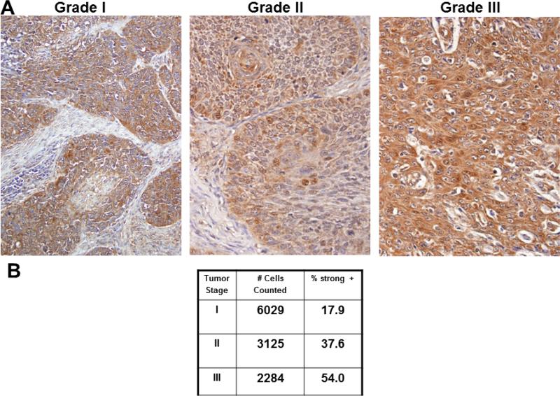
Figure 5. Immunohistochemical analysis of RelA staining in human tongue tumors.
(A) Representative image fields from staged human tongue SCC tissue microarray stained with antibodies directed against RelA. (B) Quantitation of staining as described in Materials and Methods. %strong positive is the sum of the percentage of cells staining 2+ and 3+.

