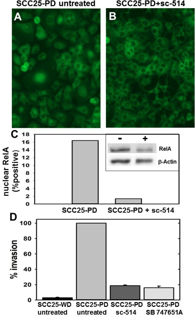
Figure 6. Inhibitors reverse nuclear translocation of RelA and block invasion.
(A,B) Cells were treated as indicated and processed for immuno-fluorescence microscopy with antibodies directed against RelA. Experiment was performed in triplicate. (A) SCC25-PD cells, untreated. (B) SCC25-PD cells treated with sc-514 (10 μM, 25 h). (C) Graph showing percentage of cells with positive nuclear RelA in control and sc-514-treated SCC25-PD cells, as indicated. (inset) Western blot of nuclear extracts of control (-) and sc-514-treated (+) SCC25-PD cells probed with antibodies directed against RelA (upper panel) and β-actin (lower panel). (D) Analysis of invasion. Cells were untreated or treated, as indicated, prior to addition to a Boyden chamber containing Matrigel as described in Experimental Procedures. Invading cells were enumerated and results shows as % of invasion relative to untreated SCC25-PD cells (designated 100%). (sc-514, 10 uM; SB 747651A, 20 uM)

