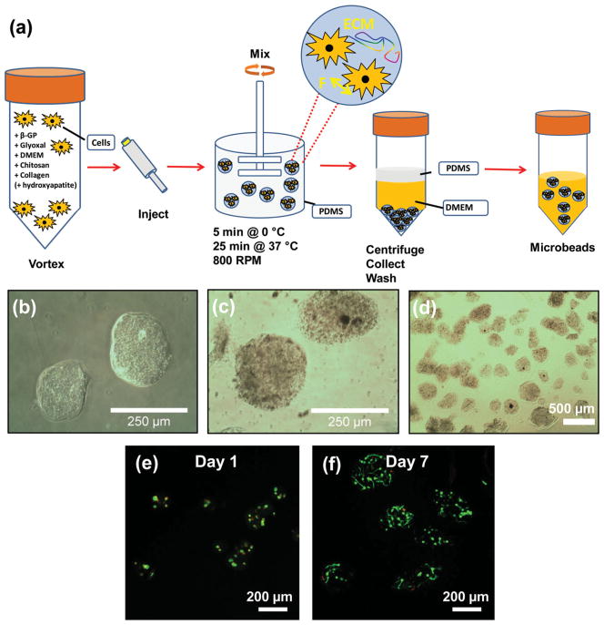
Figure 1.
a) Schematic illustration of microbead fabrication process. b, c) Light microscopy images of 65–35 wt% collagen-chitosan microbeads (panel c show microbeads containing 2.5 mg/mL hydroxyapatite). d) Light microscopy of a population of collagen/chitosan microspheres showing their generally spheroidal morphology. e, f) Fluorescence microscopy of human mesenchymal stem cells embedded in 65–35 wt% collagen-chitosan microbeads at day 1 and day 7 of culture, respectively. The cytoplasm of living cells is stained green, and the nuclei of dead cells are stained red.

