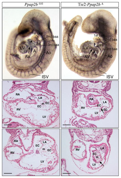
Figure 1. LPP3 is required for early vascular development.
Morphological analysis of Ppap2bfl/fl (top right panel) and Tie2-Ppap2bΔ (top left panel) embryos with 22 pairs of somites immunostained for PECAM1 and cleared with BB:BA. The Tie2-Ppap2bΔ embryo displays a smaller common atrial chamber (A) and open atrioventricular canal (AV). In addition, abnormalities of the aortic sac (as) and outflow tract (*), expansion of aorta (Ao) at the level of the 2nd branchial arch artery (baa), swelling of the 3rd bba and irregular intersomitic vasculature (ISV) were noted. Histologycal analysis (middle and bottom panels) in transverse sections of Ppap2bfl/fl (right panels) and Tie2-Ppap2bΔ (left panels) E9.5 embryos at the level of the AV canal. Note the reduction of mesenchymal (m) cells in endocardial cushions (EC) leading to a completely or partially open AV canal (arrows). LA, left component of atrial chamber; LV, left ventricle; RA, right component of atrial chamber; RV, right ventricle. Scale bar= 100 μm

