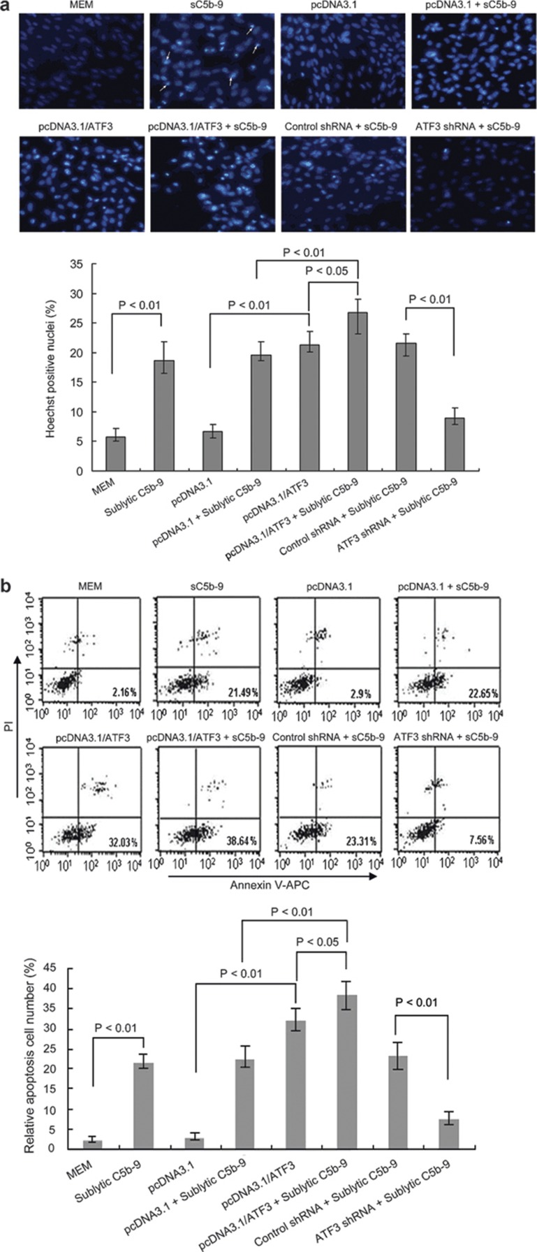
Figure 4.
ATF3 increased apoptosis of rat GMCs induced by sublytic C5b-9 (sC5b-9) attack. (a) The cultured rat GMCs were transfected with pcDNA3.1/ATF3, pcDNA3.1, shRNA, or control shRNA for 48 h and then treated with or without sublytic C5b-9 for 3 h. The cells were fixed in 3% paraformaldehyde for 30 min, washed, stained with Hoechst 33342 and examined under a fluorescence microscope. Apoptotic cells were identified as cells with condensed, disrupted nuclei (arrow, Hoechst staining, ×200). Apoptotic cells in at least five random fields were counted. The percentage of apoptotic nuclei was calculated with the following formula: 100×number of apoptotic/total counted cell number (%). At least 400 apoptotic cells were counted for the ATF3-positive staining groups. The data showed that Hoechst 33342-stained cells in the pcDNA3.1/ATF3 group or sublytic C5b-9 group were significantly higher than those in the MEM or pcDNA3.1 group, respectively (P<0.01). Meanwhile, Hoechst 33342-stained cells in the pcDNA3.1/ATF3+sublytic C5b-9 group were also higher than those in the pcDNA3.1/ATF3 (P<0.05) or pcDNA3.1+sublytic C5b-9 group (P<0.01). However, the positive cells in the ATF3 shRNA+sublytic C5b-9 group were less than that in the control shRNA+sublytic C5b-9 group (P<0.01). The result is representative of three independent experiments. (b) Differently treated GMCs were stained with Annexin V/APC and PI and analyzed by flow cytometry. Percentages of early apoptotic cells (Annexin V positive, PI negative) were calculated. In parallel, similar changes in the apoptosis percentage in the GMC groups as the above-mentioned treatments by Hoechst 33342 staining were found. The displayed result is representative of three independent experiments. APC, allophycocyanin; ATF3, activating transcription factor 3; GMC, glomerular mesangial cell; MEM, modified Eagle's medium; PI, propidium iodide; shRNA, small hairpin RNA.

