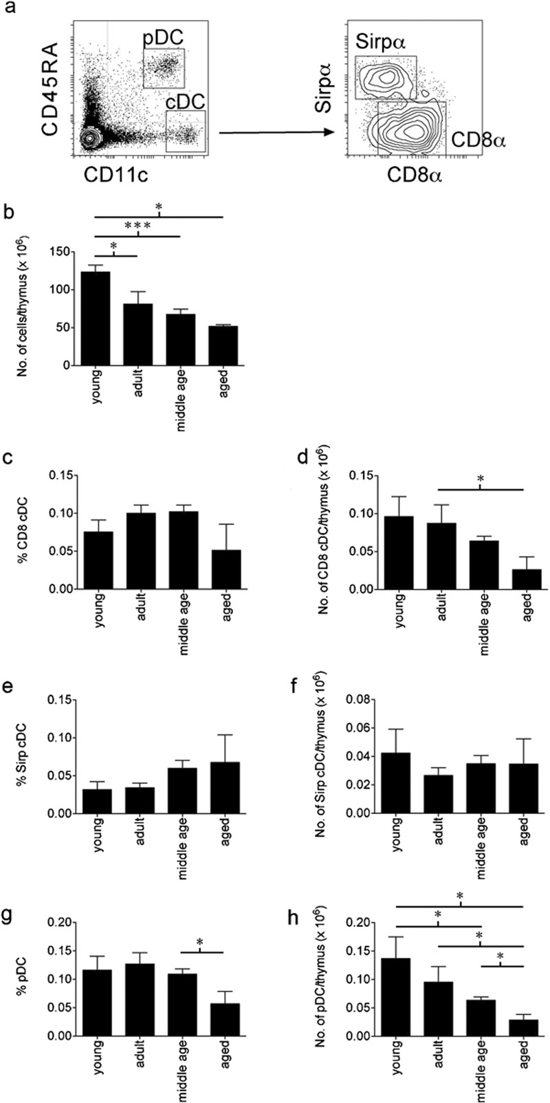
Figure 1.

Changes in the cellularity and percentages of the thymic DC subsets within the ageing thymus. (a) The thymic DC subsets were defined by flow cytometric analysis as pDC or cDC based on expression of CD11c and CD45RA. The cDC were further defined as CD8α+ cDC or Sirpα+ cDC based on expression of Sirpα or CD8α. Thymuses of young (8–12 weeks), adult (4–6 months), middle-aged (8–12 months) and aged (16–24 months) C57BL/6 mice were assessed for (b) the total number of thymocytes, (c, e, g) the percentage of each DC subset comprising the total thymocyte population and (d, f, h) the numbers of cells within each DC subset. For each age group of mice, data are shown as the pooled results of several separate experiments where n=9 for young mice, n=10 for adult mice, n=25 for middle-aged mice and n=14 for aged mice. Data are represented as mean±SEM. cDC, conventional dendritic cells; DC, dendritic cells; pDC, plasmacytoid dendritic cells; Sirpα, signal-regulatory protein-α.
