Figure 6.
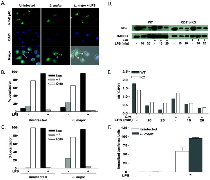
Leishmania infection does not inhibit LPS-induced NFκB activation. Representative immunofluorescence images of NFκB p65 (A) detail that infection of WT BMMP does not inhibit LPS-induced NFκB p65. WT (B) and CD11b KO (C) BMMP were seeded to glass coverslips and infected with L. major for 16 hr with or without LPS stimulation. Cells were fixed, antibody treated and DAPI stained to visualize NFκB p65 (green) and the cell nucleus (blue). Cytoplasm (C) and nuclei (N) are denoted by arrows. Nuclear (Nuc), cytoplasmic (Cyto), or intermediate (+/−) localization was quantified by counting at least 100 cells on each duplicate coverslip (B and C). Cells were collected and analyzed by Western Blot for IκB degradation (D). GAPDH was assessed as a protein loading control. Densitometry readings for IkB were normalized to GAPDH (E). One representative of three independent experiments is presented. (F) RAW 264.7 cells stably transfected with a NFκB luciferase reporter were infected with L. major for 16 hr followed by LPS stimulation for 8 hours. Samples were analyzed in triplicate. *p≤0.05 (Wilcoxon Matched Pair test; n=8) compared to unstimulated control. One representative of three independent experiments is presented.

