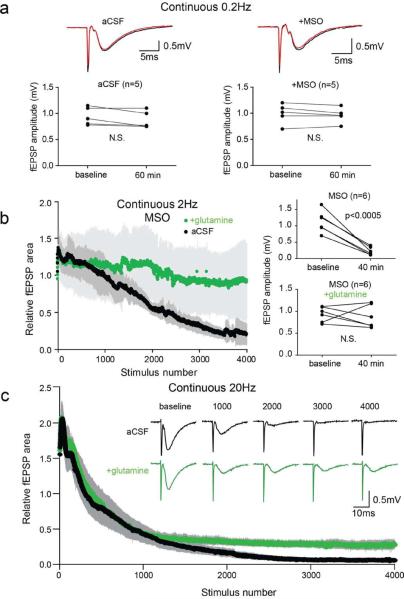
Figure 1. Glutamine prevents fEPSP depression during high frequency repetitive stimulation of the Schaffer collaterals.
(a) Example traces at baseline (black) and after 60 min (red) of continuous 0.2 Hz stimulation (upper) and paired plot of fEPSP amplitudes (lower) for untreated slices (left) and slices pretreated with MSO to inhibit glutamine synthetase (right). (b) Time course of relative fEPSP area during continuous 2 Hz stimulation (4000 stimuli; 33.3 min) of the Schaffer collaterals (left panel) in slices treated with MSO in aCSF (black; n=6) and with addition of 500 μM glutamine (green; n=6). Evoked fEPSP amplitudes during 0.2 Hz stimulation at baseline and at 40 min (~6 min after 2 Hz stimulation) shows significant fEPSP depression (Student's t-test, p<0.0005) in MSO treated slices but not in glutamine (right panel). (c) Time course of continuous 20 Hz stimulation of Schaffer collaterals in aCSF (black; n=6) and with glutamine (green, n=4). Inset: example traces at indicated stimulus numbers. Gray bars indicate SEM.

