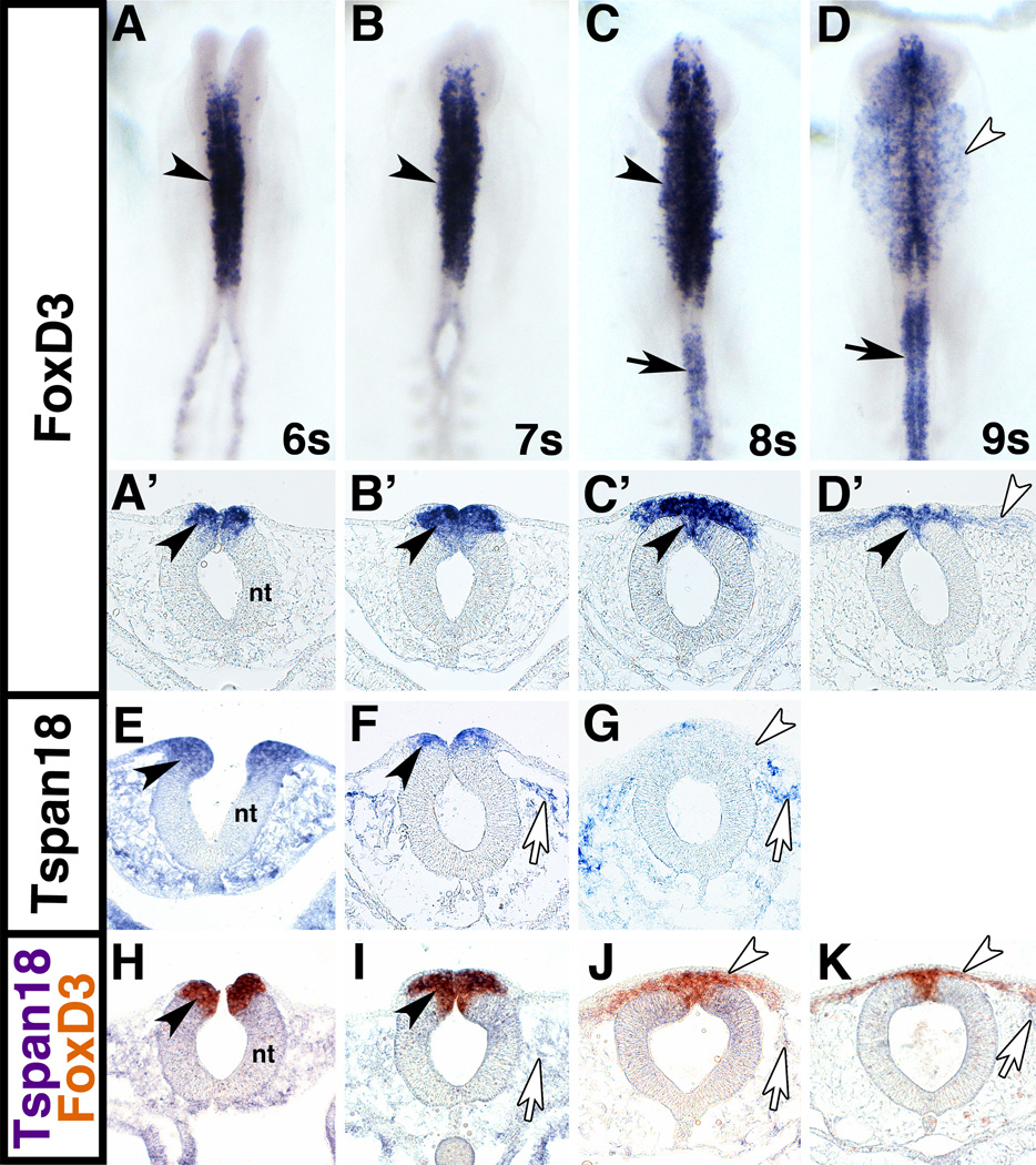
Fig. 1. FoxD3 and Tspan18 are co-expressed in premigratory cranial neural crest cells.
Whole mount and transverse sections of chick embryos processed by in situ hybridization for FoxD3 (A-D), Tspan18 (E-G), or FoxD3 and Tspan18 (H-K) at 6 somites (s; A,A’,E, H), 7s (B,B’,F, I), 8s (C,C’,G, J) and 9s (D,D’, K). (A,B) FoxD3 (purple) is expressed in premigratory cranial neural crest cells at 6 and 7s (black arrowheads). (C) At 8s, FoxD3 expression persists in early emigrating neural crest cells (black arrowheads) and extends into the trunk (black arrow). (D) At 9s, FoxD3 expression is reduced in migrating cranial neural crest cells (white arrowhead), but retained in the dorsal neural tube (black arrowhead in D’) and in the trunk (arrow). (E-G) Tspan18 (purple) is expressed in premigratory cranial neural crest cells at 6 and 7s (black arrowheads), when its expression begins to downregulate. At 8s, Tspan18 is absent from migratory neural crest cells (white arrowhead). Tspan18 is expressed in scattered cells in the head mesenchyme (white arrows). (H-K) FoxD3 (orange) and Tspan18 (purple) expression domains overlap in the neural folds at 6s and 7s (black arrowheads). At 8s and 9s, migratory neural crest cells express FoxD3 but not Tspan18 (white arrowheads); Tspan18 expression is limited to head mesenchyme (white arrows). A-D, dorsal view; A’-D’, E-K, transverse sections. nt, neural tube.

