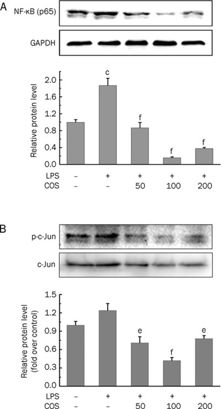
Figure 4.
Blocking effect of COS on LPS-induced NF-κB and AP-1 translocation into nucleus of HUVECs. (A) Relative protein levels of NF-κB in nucleus of HUVECs. (B) Relative protein levels of AP-1 in nucleus of HUVECs. Cells were pretreated with COS (50, 100, and 200 μg/mL) for 24 h and then exposed to LPS (100 ng/mL) for 4 h. After treatment, nuclear and cytoplasmic fractions were analyzed for detection of NF-κB by Western blot analysis as described in Materials and methods. Data are representative of three experiments (mans±SD). cP<0.01 compared to the vehicle-treated group; eP<0.05, fP<0.01 compared to the LPS-treated group.

