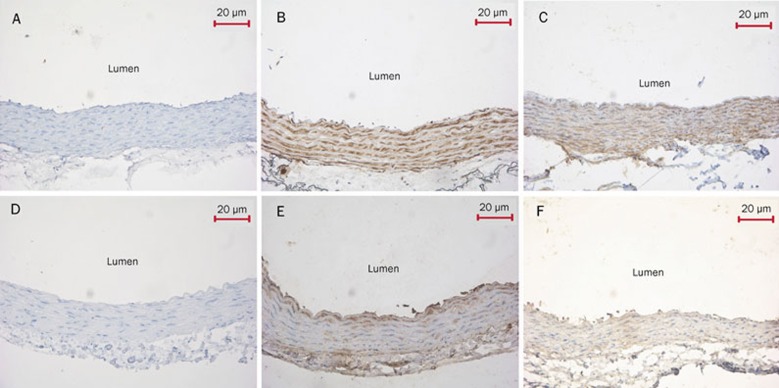
Figure 6.
Immunohistochemical analysis of SUR2B and Kir6.1 expression in aortic and pulmonary artery rings (DAB×200). (A) The aortic ring negative control was processed without SUR2B primary antibody. This control had normal smooth muscle cell and endothelial cell structure without brown granules. (B) The aortic ring was treated with SUR2B antibody. SUR2B protein was strongly expressed in the inner membrane and tunica media vasorum. The presence of the brown granules in aortic smooth muscle cells and endothelial cells was defined as positive signals. (C) The pulmonary artery ring was processed with SUR2B antibody. Brown granules were observed in pulmonary artery smooth muscle cells and endothelial cells. (D) The aortic ring negative control was treated without Kir6.1 primary antibody. The structure of smooth muscle cells and endothelial cells was normal and had no brown granules. (E) The aortic ring was treated with Kir6.1 antibody. Brown granules were strongly expressed in the medial layer of aortic ring. (F) The pulmonary artery ring was processed with Kir6.1 antibody. The brown granules were observed in pulmonary artery smooth muscle cells and endothelial cells. SUR2B, a KATP channel subunit. Kir6.1, a KATP channel subunit.

