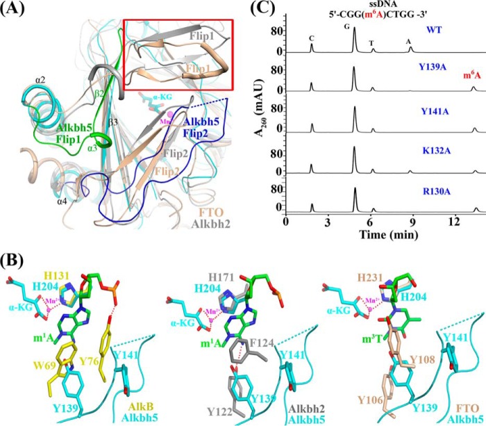
FIGURE 2.
The distinct lid region of Alkbh5 is vital for its substrate recognition and catalysis. A, structural comparison of Alkbh5 with FTO (tinted; Protein Data Bank code 3LFM) and Alkbh2 (gray; Protein Data Bank code 3BTZ) around the lid region. The most distinct regions, Flip1 (residues 117–129) and Flip2 (residues 136–165), of Alkbh5 are shown in green and blue, respectively. The discrete region is shown in blue dashed lines. The uncovered and larger space over the active site of Alkbh5 is labeled in the red frame. B, superposition of the partial key residues involved in nucleoside recognition. Alkbh5 (cyan), AlkB (Protein Data Bank code 3BIE; yellow; left panel), Alkbh2 (Protein Data Bank code 3BTZ; gray; middle panel), and FTO (Protein Data Bank code 3LFM; tinted; right panel) are shown. The nucleoside is colored green. Hydrogen bonds are indicated as red dashed lines. α-KG and Mn2+ from Alkbh5 are labeled. C, mutations of the key residues in the lid region (shown in supplemental Movie S1) greatly impair Alkbh5 demethylation activity. mAU, milli-absorbance units.

