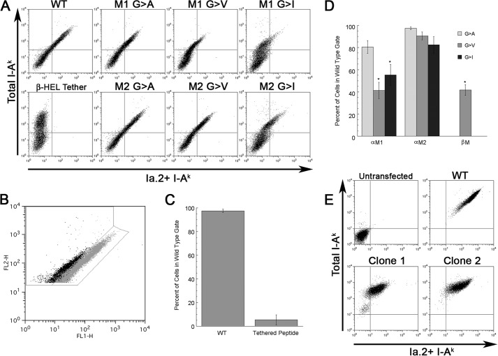
FIGURE 4.
Mutation of the I-Ak α chain M1 GXXXG motif results in selective diminution of Ia. 2+ class II expression. A, CIITA-expressing 293T cells were transfected with the indicated I-Ak α (Aαk) and β (Aβk) chain cDNAs and cultured overnight to allow protein expression. Cells were stained with anti-Ia.2 (11-5.2-FITC) and anti-Ia.17 (10-3.6-PE) and analyzed by flow cytometry (gating on single live cells by forward scatter/side scatter and lack of propidium iodide staining). Shown is the Ia.2 versus Ia.17 expression for each αβ chain pairing. Cells expressing WT I-Ak stain with both antibodies (upper right quadrant of dot plot). Cells expressing I-Ak with a tethered peptide (Aαk/Aβk-HEL) have essentially no Ia.2 staining as reported previously (5). Cells expressing α chain M1 mutations such as M1 G → V stain robustly for total class II (Ia.17) but exhibit decreased staining for Ia.2. Shown are representative results from one of greater than three independent experiments. B, gating strategy for quantification of Ia.2 epitope expression. The 11-5.2-FITC and 10-3.6-PE staining of viable class II-expressing 293T cells transfected with either WT (gray) or M1 G → V mutant (black) I-Ak class II is shown. Shown is the gate for WT Ia.2 expression as well as decreased Ia.2 expression. C and D, plots of frequency of I-Ak class II-positive transfected 293T cells that fall within the wild type gate (panel B). Error bars indicate ± 1 S.D. Reported are average values from 3–4 independent experiments. E, K46μ B cells were transfected with either WT I-Ak (WT Aαk + WT Aβk) or M1 G → A I-Ak (Aαk M1 G → A + WT Aβk) cDNAs, and stable transfectants were isolated and cloned (Clone 1 and Clone 2 are two representative clones). Cells were stained with 11-5.2-FITC and 10-3.6-PE and analyzed by flow cytometry (gating on single live cells by forward scatter/side scatter and lack of propidium iodide staining). Shown are representative results from 1 of 2 independent experiments where the same clones were stained with the 11-5.2 and 10-3.6 mAb. The two M1 G → A clones exhibit a 60% decrease in total I-Ak expression (i.e. they express 40% (41 and 39%) of the level of total class II as WT cells). If the M1 mutation had no effect on Ia.2+ conformer formation, the cells would be expected to also express 60% of the Ia.2 epitope as WT. However, the M1 mutant cells express only about 9% (6.8 and 11.6%) the level of WT Ia.2 conformer, which is ∼75% less than would be expected.

