
FIGURE 4.
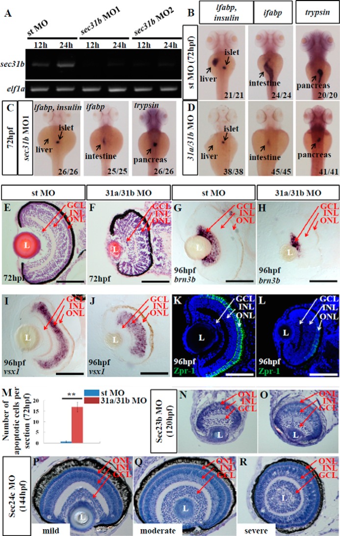
Loss-of-function of COPII does not disrupt retinal lamination. A, expression of sec31b in the embryos injected with the standard control morpholino (st MO) or two sec31b splicing morpholinos (sec31b MO1 and sec31b MO2) at 12 and 24 hpf were determined by qPCR with the expression of elf1a as the normalization control. B–D, WISH analysis of digestive organs in the control morphant (st MO), sec31b morphant, and sec31a/sec31b double morphant at 72 hpf. In each case, the number of total embryos examined (as denominator) and the number of embryos exhibiting the displayed phenotype (as numerator) are shown on the bottom right. E and F, H&E staining of the sections of eyes in the control (E) and sec31a/sec31b double (F) morphant. G–L, ganglion cells were detected with a brn3b probe (G and H), bipolar cells with a vsx1 probe (I and J), and green/red double cones with an anti-Zpr-1 antibody (K and L). M, comparison of the number of apoptotic cells in the control and sec31a/sec31b double morphant at 72 hpf (n = 3, four sections from each embryo were used for counting the apoptotic cells). N–R, histological analysis of the retinal structure in Sec23b morphant at 120 hpf and in Sec24c morphant at 144 hpf. L, lens. Scale bar, 50 μm (E–L).