
FIGURE 7.
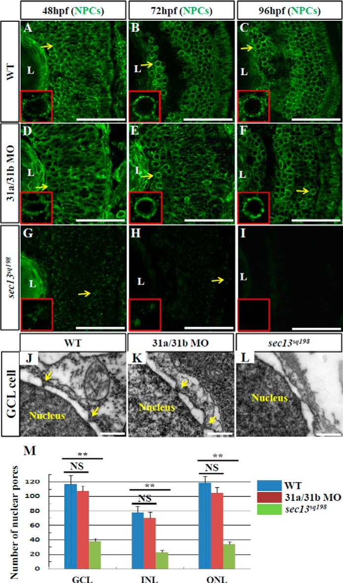
Formation of nuclear pores is impaired in the sec13sq198 mutant retina. A–I, immunostaining of nuclear pores in WT (A–C), sec131a/sec31b double morphant (D–F), and sec13sq198 mutant (G–I) retina with Mab414 antibody. The nuclear pores in both WT (A–C) and the sec31a/sec31b double morphant (D–F) retina were well formed (shown by the yellow arrows and insets), whereas formation of the nuclear pores in the sec13sq198 mutant retina gradually failed (G–I) (shown by the yellow arrows and insets). J–L, TEM analysis of the nuclear pores in the GCL cells in WT (J), sec31a/sec31b double morphant (K), and sec13sq198 mutant (L) retina at 72 hpf. Note that both the WT (J) and the sec31a/sec31b double morphant (K) retinal cells displayed characteristic nuclear pores (indicated by the yellow arrows). In contrast, the mutant retina was defective in the formation of nuclear pores in cells from all three layers. M, comparison of the number of nuclear pores in the GCL, INL, and ONL in the WT, sec31a/sec31b double morphants, and sec13sq198 mutant retina at 72 hpf. Three ultrathin sections from two embryos for each genotype were used to count the number of nuclear pores in the GCL, INL, and ONL, respectively. For each ultrathin section, six cells in each retinal layer were selected for counting, and the total number of nuclear pores from 18 cells in each retinal layer is shown here. L, lens. Scale bar, 50 μm (A–I) and 1 μm (J–L). NS, no significance. **, p < 0.01.