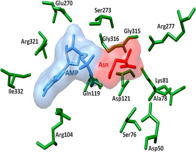
FIGURE 9.

Superposition of EcAsnA complexed with Asn and AMP and TbASNA. Ligands are shown as space-filling models. l-Asn (red), AMP (blue), and side chains of TbASNA are shown (green). The corresponding EcAsnA side chains have been removed for simplicity. All the l-Asn- and AMP-binding side chains are conserved between E. coli and TbASNA and LdASNA.
