
Figure 6.
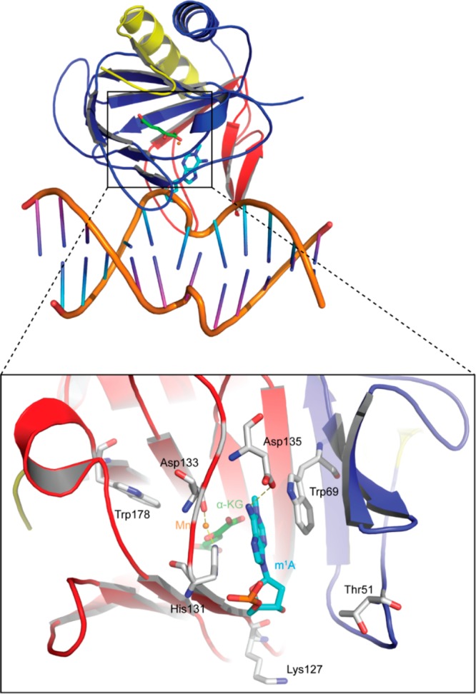
Crystal structure of an AlkB–dsDNA complex (PDB ID 3BIE). The protein is colored according to subdomain organization with the N-terminal extension in yellow (residues 13–44), the nucleotide-recognition lid in red (residues 45–90), and the catalytic core in blue (residues 91–214). Manganese(II) (orange) replaces iron(II) in the structure to eliminate catalytic activity. The flipped-base m1A is shown in blue, α-KG in green, protein residues in white, DNA backbone in beige, bases in the m1A-containing DNA strand in cyan, and bases of the complementary strand in purple.