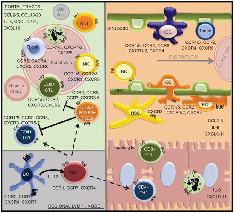
Figure 1.
Chemokine expression profile compartmentalizes the immune response to HCV in the chronically infected liver. Blood enters the liver lobule from the hepatic artery and portal vein, and flows via the sinusoids towards the central vein. Extravasation of leukocytes out of the blood and physical trapping of specific effector subsets is mediated by chemokine secretion profiles in discrete anatomical compartments. Potent cytotoxic reactions ensue within the PORTAL TRACTS retained by upregulated expression of CCR5 associated chemokines (CCL3, CCL4 and CCL5) on portal endothelium, while a nonspecific CCR5+/CXCR3+ Th1 subset dominates the helper cell population in the SINUSOIDS and hepatic parenchyma attracted by an upregulated expression of CXCL9–11. Upon maturation, dendritic cell and T-cell subtypes change surface expression of chemokine receptor CCR, facilitating their migration between secondary lymphatic tissues (REGIONAL LYMPH NODES) and the liver. CCR, CC-chemokine receptor; CTL, cytotoxic T lymphocyte; CXCR, CXC chemokine receptor; HCV, hepatitis C virus; HSC, hepatic stellate cell; iDC, immature dendritic cell; KC, Kupffer cell; LSEC, liver sinusoidal endothelial cell; NK, natural killer; PMN, polymorphonuclear neutrophil; Tmem, memory T cell; Th1, type 1 helper T cell; Treg, regulatory T cell.

