
Figure 14.
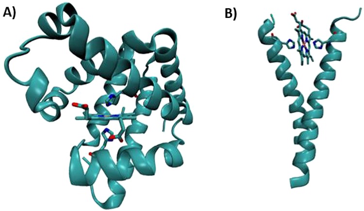
Structural models of designed cytochrome models in native scaffolds. (A) X-ray crystallographic model of a pig myoglobin designed to have cytochrome-like bis-His ligation (PDB ID 1MNI).440 (B) Molecular dynamics model of a histidine mutant of the membrane protein, glycophorin A, designed to bind heme in a cytochrome-like manner.441 Coordinates provided by courtesy of G. Ghirlanda.